A Trainee Phlebotomist Caused a Hematoma
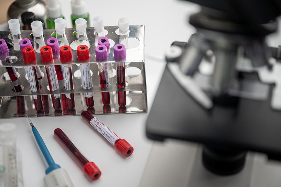
Hematoma: Understanding the Bruising Beneath the Skin
From the smallest bump to the most severe trauma, our bodies are remarkably adept at healing themselves. One of the body’s remarkable defense mechanisms against injury is the formation of a hematoma. But what exactly is a hematoma, and how does it play a vital role in the healing process?
Defining Hematoma
A hematoma is a localized collection of blood that pools outside the blood vessels, usually within the soft tissues of the body, such as muscles or skin. Essentially, it is a bruise beneath the skin’s surface. Hematomas can range in size from small and inconspicuous to large and painful. They typically occur as a result of trauma, injury, or surgery but can also develop spontaneously in some medical conditions.
The Mechanism of Hematoma Formation
When a blood vessel, such as an artery, vein, or capillary, is damaged due to injury, the body activates a series of processes to stop bleeding and repair the vessel. This process is known as hemostasis.
- Vasoconstriction: The initial response to vessel damage is vasoconstriction, where the blood vessel narrows to reduce blood flow to the injured area. This helps minimize blood loss.
- Platelet Activation: Platelets, small cell fragments in the bloodstream, play a crucial role in forming blood clots. They quickly adhere to the damaged vessel wall, creating a temporary plug to stop bleeding.
- Coagulation Cascade: A complex sequence of events known as the coagulation cascade is initiated. This process involves the activation of various clotting factors in the blood, eventually leading to the formation of a stable blood clot or thrombus.
- Clot Retraction: After the initial clot forms, it begins to contract or retract, pulling the edges of the damaged blood vessel together. This helps seal the vessel and prevent further bleeding.
- Fibrin Formation: Fibrin, a protein, forms a meshwork within the clot, further strengthening it.
Despite these intricate mechanisms, in some cases, blood can still leak out of the damaged vessel and accumulate in the surrounding tissues, leading to the formation of a hematoma.
Types of Hematomas
Hematomas can be classified into several types based on their location and the underlying cause:
- Subcutaneous Hematoma: These occur beneath the skin and are often visible as a bruise or discoloration. They are commonly caused by blunt trauma.
- Intramuscular Hematoma: These form within muscles and are typically associated with muscle injuries or strenuous physical activity.
- Subdural Hematoma: These are located between the brain and the protective covering (the dura) and often result from head injuries.
- Epidural Hematoma: These occur between the skull and the outermost layer of the brain’s protective covering (the dura) and are usually caused by severe head trauma.
- Intracranial Hematoma: These hematomas form within the brain tissue and can result from various causes, including head injuries or blood vessel abnormalities.
Treatment and Management
The management of a hematoma depends on its location, size, and the underlying cause. In many cases, small hematomas resolve on their own as the body reabsorbs the trapped blood. However, larger or more severe hematomas may require medical intervention.
Treatment options may include:
- Rest and Elevation: Elevating the affected area and resting can help reduce swelling and improve blood circulation.
- Cold Compresses: Applying cold compresses or ice packs to the area can help reduce pain and inflammation.
- Compression: In some cases, compression bandages may be recommended to prevent further bleeding and reduce swelling.
- Surgical Drainage: For large or deep hematomas, surgical drainage may be necessary to remove the accumulated blood and relieve pressure on surrounding tissues.
- Medications: In cases where blood clotting is impaired, or to prevent complications like infection, medication may be prescribed.
Medical Negligence: A Trainee Phlebotomist Caused A Hematoma
A hematoma is a common occurrence in the field of phlebotomy, and it can sometimes be an unintended outcome of the blood collection process. A trainee phlebotomist, while learning and perfecting their skills, may inadvertently cause a hematoma. Let’s explore how this can happen and what steps can be taken to minimize the risk of hematoma formation during blood collection.
- Needle Insertion Technique: One of the key factors in preventing hematomas is the proper technique for needle insertion. A trainee phlebotomist may sometimes insert the needle too deeply, causing damage to not only the vein but also the surrounding tissues. This can lead to blood leaking out of the vein and into the surrounding tissue, resulting in a hematoma.
- Vein Selection: Choosing the right vein is crucial. A trainee phlebotomist might select a vein that is too small, fragile, or prone to rolling. This can make it more challenging to successfully access the vein and increase the risk of hematoma formation.
- Needle Positioning: The angle at which the needle is inserted matters. If the needle is not inserted at the correct angle, it can increase the likelihood of vein puncture and hematoma.
- Failure to Release Tourniquet: Leaving the tourniquet on for an extended period can cause blood to pool in the vein, increasing the risk of hematoma when the tourniquet is finally released.
- Improper Pressure Application: After withdrawing the needle, it’s essential to apply adequate pressure to the puncture site to help prevent blood from leaking into the surrounding tissue. A trainee phlebotomist may not apply sufficient pressure, leading to hematoma formation.
- Inadequate Observation: Monitoring the patient for signs of hematoma during and after the procedure is crucial. A trainee phlebotomist may not be experienced enough to recognize the early signs of a hematoma developing.
Minimizing the Risk of Hematomas in Phlebotomy:
To reduce the risk of hematoma formation during blood collection, trainee phlebotomists should follow these best practices:
- Proper Training: Thorough training and supervised practice are essential for phlebotomy students. This includes learning proper techniques for needle insertion, vein selection, and patient care.
- Vein Assessment: Before attempting venipuncture, assess the patient’s veins carefully. Choose a suitable vein that is visible, palpable, and less likely to roll.
- Correct Needle Angle: Ensure that the needle is inserted at the appropriate angle (usually around 15-30 degrees) to minimize the risk of vein damage.
- Tourniquet Control: Use the tourniquet judiciously, and release it promptly once the vein is accessed and blood begins to flow.
- Proper Pressure Application: After needle removal, apply gentle pressure to the puncture site with gauze or a cotton ball, holding it in place for a sufficient amount of time to allow the vein to seal.
- Patient Education: Communicate with the patient throughout the process, explaining what is happening and what they can expect. Reducing anxiety can help minimize patient movement, which can contribute to hematoma formation.
- Post-Procedure Observation: After blood collection, monitor the puncture site for any signs of hematoma formation and take appropriate action if necessary.
Understanding the Risks of Thrombotic Thrombocytopenic Purpura (TTP) and Hematoma
Thrombotic Thrombocytopenic Purpura (TTP) and hematoma are distinct medical conditions, each with its own set of risks and implications. However, they can be interconnected in certain scenarios, especially when TTP affects the blood’s ability to clot properly. Let’s explore these two conditions and their associated risks.
Thrombotic Thrombocytopenic Purpura (TTP):
TTP is a rare and potentially life-threatening blood disorder characterized by the formation of small blood clots throughout the body. It primarily affects the smallest blood vessels, leading to a cascade of health issues. TTP is caused by a deficiency of a specific enzyme called ADAMTS13, which is responsible for breaking down a large protein called von Willebrand factor (vWF). Without sufficient ADAMTS13, vWF accumulates, leading to abnormal blood clot formation.
Risks of TTP:
- Clot Formation: The hallmark of TTP is the formation of microclots in small blood vessels throughout the body. This can lead to organ damage and dysfunction, as the clots impede blood flow to vital organs like the brain, kidneys, and heart.
- Bleeding Tendency: Paradoxically, even though TTP is characterized by clot formation, it can also cause a decrease in platelet count (thrombocytopenia), leading to an increased risk of bleeding. This dual risk of clotting and bleeding makes TTP a challenging condition to manage.
- Organ Damage: When clots disrupt blood flow to organs, it can result in damage and failure. For example, TTP-induced clots in the brain can cause strokes, while those in the kidneys can lead to kidney dysfunction.
- Neurological Complications: TTP can lead to neurological symptoms, including confusion, weakness, and even coma, due to the impact of clots on the brain.
Risks of Hematoma:
- Pain and Swelling: Hematomas can cause significant pain and swelling at the site of the injury. This can interfere with the function of the affected body part and disrupt daily activities.
- Infection: There is a risk of infection if the hematoma breaks through the skin or if the injury that caused the hematoma is contaminated.
- Compartment Syndrome: In severe cases, a hematoma can cause increased pressure within the affected area, known as compartment syndrome. This can lead to tissue damage and impaired blood flow, necessitating immediate medical attention.
- Secondary Complications: Depending on the location and size of the hematoma, it can lead to secondary complications. For example, a hematoma near a joint may result in limited mobility.
The Connection Between TTP and Hematoma:
While TTP primarily involves abnormal clot formation within the bloodstream, it can impact the body’s overall ability to regulate clotting and bleeding. In some cases, individuals with TTP may be at an increased risk of both bleeding and clotting disorders. This dual risk might predispose them to the formation of hematomas, particularly if they experience trauma or injury.
Managing TTP typically involves therapies to suppress abnormal clot formation, such as plasma exchange and medication. In individuals with TTP, healthcare providers must carefully assess and address the risks of both bleeding and clotting to provide effective treatment.
Immune Thrombocytopenia (ITP)
ITP, or Immune Thrombocytopenia, is a medical condition characterized by a low platelet count in the blood. Platelets are a type of blood cell that plays a crucial role in blood clotting, and a low platelet count can lead to bleeding and easy bruising.
In ITP, the immune system mistakenly targets and destroys platelets as if they were foreign invaders. This is typically due to an autoimmune response, where the body’s immune system attacks its own healthy cells and tissues. In the case of ITP, it is believed that antibodies produced by the immune system bind to platelets, marking them for destruction by the spleen or other immune cells.
The exact cause of ITP is not always clear, but it is often considered an autoimmune disorder. It can occur in isolation, known as primary or idiopathic ITP, where there is no underlying cause, or it can be secondary to other autoimmune disorders or conditions such as lupus, rheumatoid arthritis, or certain infections.
The symptoms of ITP can vary from mild to severe and may include:
- Easy bruising.
- Prolonged bleeding from small cuts.
- Petechiae (small red or purple spots on the skin).
- Nosebleeds.
- Blood in the urine or stools.
- Heavy menstrual periods.
- In severe cases, internal bleeding can be life-threatening.
Treatment for ITP may vary depending on the severity of the condition. Some individuals with mild ITP may not require treatment and can be managed through regular monitoring. In more severe cases or when bleeding symptoms are significant, treatment options may include:
- Corticosteroids to suppress the immune response.
- Intravenous immunoglobulin (IVIG) to temporarily raise platelet counts.
- Platelet transfusions in cases of severe bleeding.
- Medications that affect the immune system, such as rituximab or thrombopoietin receptor agonists.
- In extreme cases, surgery to remove the spleen (splenectomy) may be considered.
Managing ITP with an underlying autoimmune disorder may involve treating the primary autoimmune condition as well. It’s essential for individuals with ITP to work closely with their healthcare team to determine the most appropriate treatment plan based on their specific circumstances and symptoms.
Both ITP (Immune Thrombocytopenia) and hematomas involve issues related to blood clotting and can carry certain risks:
Risks of ITP (Immune Thrombocytopenia):
- Bleeding: The primary risk associated with ITP is bleeding due to a low platelet count. Platelets are essential for normal blood clotting, and when their numbers are low in ITP, there’s an increased risk of spontaneous bleeding, such as nosebleeds, gum bleeding, easy bruising, and, in severe cases, internal bleeding.
- Severe Bleeding Episodes: In severe cases of ITP, there is a risk of life-threatening bleeding, such as gastrointestinal bleeding or intracranial hemorrhage. These are medical emergencies and require immediate attention.
- Chronic Disease: ITP can become a chronic condition for some individuals, which means they may experience ongoing issues with low platelet counts and the associated risk of bleeding over an extended period.
- Impact on Quality of Life: Living with ITP can be challenging due to the need for frequent medical monitoring, potential treatment side effects, and lifestyle adjustments to reduce the risk of bleeding.
The risks associated with ITP and hematomas can vary depending on the severity of the condition, the individual’s overall health, and the location and size of the hematoma. It’s essential to seek medical attention and follow medical advice for both conditions to manage and reduce these risks effectively. For ITP, this may involve treatments to increase platelet counts or manage bleeding, while for hematomas, treatment may include drainage, wound care, or surgical intervention if necessary.
Personal Experience
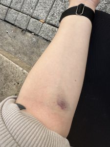
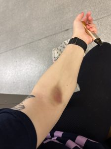
As a multiple sclerosis sufferer who has had Alemtuzumab Treatment with regular blood tests required by law, upon visiting the hospital on Wednesday 20th September 2023 I was greeted by a trainee phlebotomist whom may I add was supervised. I did not take either of their names. I explained I have problems with my veins being too thin, The trained phlebotomist looked at my arm and told the trainee phlebotomist to try the fatter vein, after a bit of poking and prodding the needle the trainee nurse took out the needle after several times going in a different direction and with a bit of perseverance she managed to draw blood. When she finally took the needle out this resulted in my arm bruising and swelling to the point it was extremely painful.
If the test tube is not removed when the needle comes out, it can cause a number of issues. For instance, the blood sample may clot inside the tube, making it difficult to extract the required amount of blood for testing. In some cases, if the needle is withdrawn too quickly, it can cause a hematoma. Hematomas can be painful and may require medical attention if they are large or do not resolve on their own.
I did have an independent nurse and pharmacist look at it afterward (not from the hospital) and both agreed it was really bad.

I realize the hospital (UHW) is a teaching hospital but the departments should get the patient’s permission first before letting trainee doctors/nurses and phlebotomists train on a patient. I was never informed until after the fact and by that time it was too late. Had I known from the start a trainee phlebotomist would be taking my blood I would have refused. This is not the first time I have had a hematoma.
My arm a week later is still bruised and swollen. This is a lesson learned. I would like to add I do not hold a grievance to the trainee phlebotomist but with my history of blood tests proving difficult, I would expect a professional fully trained nurse to take my blood.
Conclusion
Hematomas are a natural part of the body’s response to injury and play a crucial role in the healing process. While they can be painful and concerning, most hematomas resolve with time and appropriate care. However, it is essential to seek medical attention if you suspect a severe hematoma, especially if it is associated with head trauma, as prompt treatment may be necessary to prevent complications. Understanding how hematomas form and the importance of their role in the healing process can help individuals make informed decisions about their care when faced with these common injuries.
It’s important to remember that hematoma formation is a potential risk in phlebotomy, even for experienced practitioners. Trainee phlebotomists should focus on learning and consistently applying proper techniques to minimize this risk and ensure safe and adequate blood collection procedures. Supervision and feedback from experienced phlebotomists are valuable tools in the learning process.
TTP and hematoma are distinct medical conditions, each associated with its own set of risks and complications. However, in some cases, individuals with TTP may be more susceptible to hematoma formation due to disruptions in their blood clotting and bleeding mechanisms. It is essential for healthcare professionals to consider these risks when managing patients with TTP and tailor treatment accordingly.
What should a patient do if a trainee phlebotomist causes a hematoma in high-risk patients who are susceptible to ITP having blood tests because of Lemtrada alemtuzumab treatment?
If a trainee phlebotomist causes a hematoma in a high-risk patient who is susceptible to ITP (Immune Thrombocytopenia) due to Lemtrada (alemtuzumab) treatment, it is crucial for the patient to take the following steps:
- Notify the Phlebotomist: Immediately inform the phlebotomist about the hematoma and any discomfort or pain you may be experiencing. They need to be aware of the issue to provide assistance and documentation.
- Apply Pressure: If the hematoma is actively bleeding or swelling, gently apply pressure to the area with a clean cloth or sterile gauze to help control bleeding and minimize the size of the hematoma. Do not press too hard, as this could exacerbate the issue.
- Elevate the Affected Area: If possible, keep the affected limb elevated. Elevating the area can help reduce swelling and minimize further blood accumulation in the hematoma.
- Seek Medical Attention: Given the patient’s susceptibility to ITP and the potential complications associated with hematoma, it is advisable to seek immediate medical attention. This is especially important because ITP is a condition characterized by abnormal clot formation and bleeding tendencies, and it could be exacerbated by a hematoma.
- Notify Healthcare Provider: Contact the healthcare provider responsible for managing your Lemtrada treatment. Inform them of the situation, including details about the hematoma and any symptoms you are experiencing. They may need to adjust your treatment plan or monitor your condition closely.
- Follow Medical Advice: Follow the guidance provided by your healthcare provider regarding the management of the hematoma and any potential treatment adjustments necessary to mitigate the risk of ITP.
- Document the Incident: Keep detailed records of the hematoma incident, including the date, time, and name of the phlebotomist involved. This documentation may be valuable for future reference, especially if it leads to complications related to your ITP susceptibility or Lemtrada treatment.
- Consider Future Precautions: Discuss with your healthcare provider and the phlebotomy department whether any additional precautions or modifications to the blood collection process are necessary to minimize the risk of hematoma formation during future blood tests. This could include using experienced phlebotomists or employing alternative blood collection methods.
It is essential to prioritize your health and safety in this situation, especially if you are a high-risk patient with ITP susceptibility due to Lemtrada treatment. Prompt and effective communication with healthcare providers is critical to ensure that any potential complications are addressed promptly, and your treatment plan is adjusted as needed.
Photographic Evidence


Further Reading
Lemtrada (alemtuzumab) | MS Trust
Relapsing MS Infusion Treatment: LEMTRADA® (alemtuzumab)
Immune Thrombocytopenic Purpura (ITP) | Patient
Thrombotic Thrombocytopenic Purpura (also known as ‘TTP’) | CUH
ADVERTISEMENT

#hematoma #blotclot #phlebotomist #traineephlebotomist #medicalnegligence #nhs #thromboticthrombocytopenicpurpura #ttp #lemtrada #multiplesclerosis #ms #msbloodtests
Many users prefer to remain anonymous online for a variety of compelling reasons. Anonymity provides a shield against potential privacy breaches, cyberbullying, or harassment, creating a safer digital environment. It also allows individuals to express themselves freely without fear of repercussions, enabling honest discussions on sensitive topics and fostering diverse perspectives. Furthermore, some users may have legitimate concerns about government surveillance or data mining by corporations, driving them to safeguard their identity. In essence, anonymity empowers individuals to engage in open discourse, explore their interests, and protect their online presence, preserving the internet as a space for free expression and diverse voices.